Contact Us
Office Hours
Alternating Saturdays
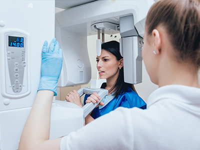
Digital radiography replaces traditional film with electronic sensors and computer processing to capture dental images. Instead of developing film in a darkroom, sensors record X-ray information and translate it into images that appear on a monitor within seconds. This transition to digital workflows has reshaped how clinicians evaluate teeth, bone, and supporting structures while making the entire imaging process faster and more efficient for patients.
There are several types of digital imaging used in contemporary dental offices, from intraoral bitewing and periapical sensors to extraoral panoramic and cone-beam systems for more complex cases. Each tool serves a different diagnostic purpose, but they share the same basic advantage: rapid capture and immediate access to images. This immediacy allows clinicians to review findings with patients and develop treatment plans without delay.
At the office in Herndon, the move to digital radiography is part of a broader commitment to precise, patient-centered care. By integrating sensors, imaging software, and secure storage systems, the team can deliver clear diagnostic information while keeping patient comfort and safety at the forefront.
One of the most frequently cited benefits of digital radiography is reduced radiation exposure compared with conventional film techniques. Modern sensors are more sensitive to X-rays, which means they can produce diagnostic-quality images with lower doses. Clinicians still follow established safety principles — using appropriate shielding, collimation, and exposure settings tailored to each patient — to keep radiation levels as low as reasonably achievable.
Because digital systems provide immediate image feedback, the need for repeat exposures is minimized. If an initial image shows suboptimal positioning or motion blur, the image can be taken again right away with adjustments, rather than reprocessing film and hoping for better results. This real-time correction capability reduces cumulative exposure over the course of a patient’s care.
Digital imaging also limits environmental hazards associated with film development. Film processing requires chemicals and physical handling; digital images eliminate that step, removing a source of workplace waste and simplifying infection-control workflows in the operatory.
Digital radiography offers more than just speed and lower dose — it provides a suite of tools that enhance diagnostic clarity. Software features such as adjustable contrast and brightness, magnification, and measurement overlays let clinicians inspect subtle details that might be harder to see on film. These enhancements are particularly useful when evaluating early cavities, fine root fractures, or small changes in bone around teeth and implants.
Because images can be instantly enlarged and manipulated on-screen, clinicians can examine problem areas from multiple visual angles without subjecting the patient to additional exposures. The ability to annotate and compare images side-by-side is also valuable when monitoring progressive conditions like periodontal bone loss or assessing the success of previous treatments.
For restorative and endodontic procedures, clear digital images help guide precise work. High-resolution intraoral images support accurate cavity detection and margin assessment for crowns and fillings, while targeted periapical views assist with root canal diagnosis and follow-up. The diagnostic flexibility of digital imaging helps clinicians make informed, timely decisions.
Beyond diagnosis, many practices use digital imaging as a visual aid during patient consultations. When patients can see their own images on a screen, it fosters clearer communication about treatment needs and expected outcomes, supporting better-informed consent and a stronger partnership in care planning.
From a patient’s perspective, the differences between film and digital imaging are noticeable. Digital sensors are often thinner and more ergonomically shaped than film holders, which can reduce discomfort during placement. Shorter exposure times and the ability to retake images quickly when necessary mean less time spent holding positions and sitting in the chair.
Because the clinician can pull images up on a monitor immediately after capture, there is typically less waiting involved. This speed contributes to shorter visits and a more streamlined workflow — particularly helpful for anxious patients, parents with young children, and those with busy schedules. Immediate visualization also enables the clinician to address concerns in real time and show patients exactly what was observed.
Infection-control practices are also simplified with digital workflows. Since there’s no need to handle exposed film or process chemicals, the operatory has fewer steps that require additional handling, which supports a cleaner, more efficient environment for both patients and staff.
Once an image is captured digitally, it can be stored securely within the practice’s patient records and retrieved instantly whenever needed. Modern dental software systems use standardized file formats and secure protocols to protect patient privacy while ensuring that images remain accessible for long-term monitoring and comparison. Secure storage reduces the risk of lost films and simplifies recordkeeping for ongoing care.
Digital files can also be shared quickly and reliably with specialists, labs, or referring providers when collaborative care is required. Electronic transfer of images speeds up consultations and makes it easier for multiple practitioners to review the same data set without the delays inherent to physical film delivery. This collaboration supports coordinated treatment planning and can accelerate decision-making for complex cases.
For practices focused on continuity of care, digital radiography enables effective tracking of a patient’s dental history over time. Longitudinal comparisons are straightforward: past and current images can be displayed together to highlight changes and measure response to treatment. That historical perspective is valuable for preventive care, chronic condition management, and long-term outcome assessment.
All of these capabilities tie back into a practice’s commitment to delivering quality care efficiently and transparently. Digital imaging is one component among many that helps clinicians provide accurate diagnoses and facilitate clear communication across the care team.
Summary: Digital radiography is a practical, patient-friendly technology that enhances diagnostic accuracy, reduces radiation exposure, and streamlines clinical workflows. By combining sensitive sensors with image-enhancement tools and secure digital storage, modern dental imaging supports faster decisions and clearer communication between clinicians and patients. If you would like to learn more about how digital radiography is used in our Herndon office, contact SmileLine Dental for more information.
Digital radiography is a dental imaging method that uses electronic sensors and computer software to capture and display X-ray images almost instantly. The sensors record X-ray information and convert it into digital images that clinicians can view on a monitor within seconds. This technology replaces traditional film and integrates with practice management systems for efficient storage and retrieval.
Digital images can be enhanced, magnified, and measured with software tools that aid clinical interpretation. The rapid feedback helps clinicians explain findings to patients and make treatment decisions during the same visit. Overall, digital radiography streamlines diagnostic workflows while preserving detailed anatomical information.
Unlike traditional film, digital radiography uses electronic sensors instead of chemical film that requires developing. Digital systems provide immediate images, eliminate the need for film processing chemicals, and reduce the physical handling steps associated with conventional film. This transition reduces environmental waste and simplifies infection-control procedures in the operatory.
Digital workflows also offer flexible image manipulation such as contrast adjustment, zoom, and measurement overlays that are not possible with unenhanced film. These enhancements can make subtle problems easier to detect and document, improving the accuracy of diagnoses and long-term monitoring. Clinicians can compare current and prior images side-by-side without waiting for film retrieval or reprocessing.
Digital radiography generally exposes patients to lower levels of radiation compared with conventional film because modern sensors are more sensitive and require smaller doses to produce diagnostic images. Clinicians adhere to established safety practices such as appropriate shielding, collimation, and exposure settings tailored to each patient to keep radiation doses as low as reasonably achievable. At SmileLine Dental in Herndon, the team follows these principles to prioritize patient safety during all imaging procedures.
Another safety advantage of digital systems is the ability to review images instantly and retake any that show positioning errors, reducing the chance of repeated exposures over time. Combined with routine equipment maintenance and adherence to recommended imaging protocols, digital radiography supports safe diagnostic imaging for patients of all ages. If you have specific radiation concerns, your clinician can explain how exposure is minimized and when imaging is clinically indicated.
Common digital imaging modalities in dental practice include intraoral bitewing and periapical sensors, extraoral panoramic radiography, and cone-beam computed tomography (CBCT) for three-dimensional imaging. Intraoral sensors are typically used for routine cavity detection and root assessments, while panoramic images capture the entire jaws and dentition in a single view. CBCT provides detailed 3D views used for implant planning, complex endodontic cases, and assessment of anatomical structures.
Each modality serves a distinct diagnostic purpose and is selected based on the clinical question at hand. The choice of imaging depends on factors such as the area of interest, diagnostic requirements, and patient anatomy. Clinicians combine these tools as needed to form a comprehensive evaluation without unnecessary exposures.
Digital radiography improves diagnosis by providing high-resolution images and software tools that enhance visualization of small details such as early caries, root fractures, or marginal gaps around restorations. Adjustable contrast, magnification, and measurement overlays allow clinicians to inspect areas that might be difficult to evaluate on film. These capabilities support more confident clinical judgments and can reveal conditions at an earlier stage.
For treatment planning, instant image access enables clinicians to correlate clinical findings with radiographic evidence during the same appointment. This immediacy helps in developing precise restorative, endodontic, or surgical plans and in explaining treatment options to patients. When needed, clinicians can annotate images and save them in the record for long-term monitoring and interdisciplinary collaboration.
Yes. Because digital images appear immediately on-screen, clinicians can verify positioning and image quality right after capture and retake any suboptimal images while the patient is still prepared. This real-time feedback reduces the likelihood of having to call a patient back for additional exposures or reprocessing. The ability to correct positioning and technique on the spot contributes to a more efficient and patient-friendly experience.
Improved sensor sensitivity also means exposures are shorter and less demanding, reducing motion blur and the frequency of retakes. Together with staff training and standardized imaging protocols, digital radiography helps minimize cumulative exposure while maintaining diagnostic confidence. Ongoing quality assurance practices further reduce repeat imaging over time.
Digital sensors are often thinner and more ergonomically shaped than traditional film holders, which can make intraoral positioning more comfortable for many patients. Shorter exposure times and fewer retakes also reduce the time a patient needs to hold intraoral devices in place. These factors are especially helpful for anxious patients, children, and those with limited tolerance for intraoral procedures.
Clinicians can also use extraoral options such as panoramic or CBCT imaging when appropriate, avoiding intraoral placement altogether for certain diagnostic needs. Clear communication about what to expect during imaging and gentle positioning techniques further enhance patient comfort. Overall, digital workflows are designed to be quicker and less intrusive without compromising diagnostic quality.
Digital images are stored in the practice management system or a secure imaging server using standardized file formats and access controls that protect patient privacy. Modern dental software typically includes user authentication, role-based permissions, and encrypted storage or transmission to reduce the risk of unauthorized access. Regular backups and IT security practices help ensure that records remain available and intact for continuity of care.
Clinicians can retrieve historical images instantly for comparison and long-term monitoring, which simplifies recordkeeping and treatment tracking. When images must be shared with other providers, secure electronic transfer methods are used to maintain privacy and data integrity. These protections are part of routine information governance in dental practices.
Many dentists use digital radiography as part of routine diagnostic care, but the need for imaging during a checkup is determined by individual risk factors, clinical findings, and screening guidelines. Clinicians assess each patient's oral health history, clinical exam results, and susceptibility to disease before recommending any radiographs. When images are clinically indicated, digital methods are often preferred for their speed and diagnostic advantages.
For patients with no symptoms and low risk, radiographic intervals may be extended according to professional guidelines and clinical judgment. Conversely, patients with active disease, restorative needs, or changes in symptoms may require more frequent imaging. Your dentist can explain the rationale for any recommended radiographs and discuss how digital imaging benefits the diagnostic process.
Digital radiography facilitates timely collaboration by enabling quick, secure sharing of images with specialists, labs, or referring providers. Electronic transfer of standardized image files accelerates consultations and allows multiple clinicians to review the same high-quality data set without delays associated with physical film. This streamlined communication supports coordinated treatment planning for complex restorative, surgical, or orthodontic cases.
Having digital images in the patient record also helps the lead clinician document findings, track progress, and provide specialists with the precise views needed for their assessment. When combined with clinical notes and intraoral photography, digital radiographs contribute to a comprehensive data set that enhances planning, reduces misunderstandings, and promotes predictable outcomes.